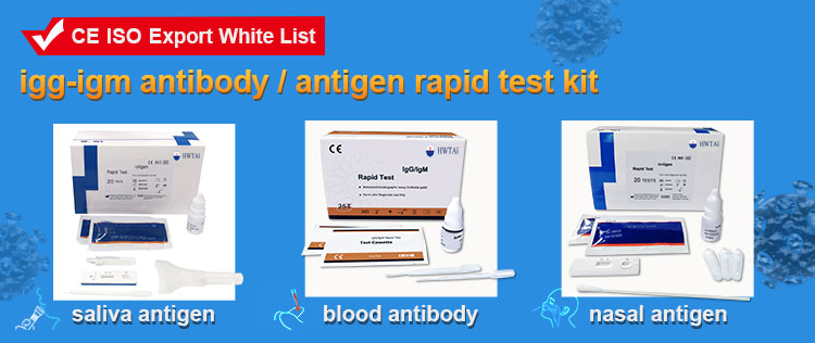
This product is used for in vitro qualitative detection of the IgG/IgM of Monkeypox in human Sliva .
CE Certificated .


Introduction
The Monkeypox virus IgG/IgM Rapid test is a rapid chromatographic immunoassay for the qualitative detection of IgG and IgM antibodies to Monkeypox virus in human whole blood, serum, or plasma as an aid in the diagnosis of Monkeypox infections.
Benefits
• Rapid testing for Monkeypox antigen within 15 minutes
• Facilitates patient treatment decisions quickly
• Simple, time-saving procedure
• All necessary reagents provided & no equipment needed
• High sensitivity and specificity
Principle
The Monkeypox virus IgG/IgM Rapid test is a qualitative membrane-based immunoassay for the detection of Monkeypox antibodies in whole blood, serum, or plasma. This test consists of two components, an IgG component and an IgM component. In the IgG component, anti-human IgG is coated in test line region G of the test. During testing, the specimen reacts with Monkeypox antigen-coated particles in the test strip. The mixture then migrates upward on the membrane chromatographically by capillary action and reacts with the anti-human IgG in test line region G. If the specimen contains IgG antibodies to Monkeypox, a colored line will appear in test line region G. In the IgM component, anti-ligand is coated in test line region M of the test. During testing, the specimen reacts with ligand anti-human IgM. Monkeypox IgM antibodies, if present in the specimen, reacts with the ligand anti-human IgM and the Monkeypox antigen-coated particles in the test strip, and this complex is captured by the anti-ligand, forming a colored line in test line region M. Therefore, if the specimen contains Monkeypox IgG antibodies, a colored line will appear in test line region G. If the specimen contains Monkeypox IgM antibodies, a colored line will appear in test line region M. If the specimen does not contain Monkeypox antibodies, no colored line will appear in either of the test line regions, indicating a negative result. To serve as a procedural control, a colored line will always change from blue to red in the control line region, indicating that theproper volume of specimen has been added and membrane wicking has occurred.
Components
• Test devices
• Droppers
• Single Buffer
• Package insert
Procedure
Allow the test device, specimen, buffer, and/or controls to reach room temperature (15-30°C) prior to testing.
1.Bring the pouch to room temperature before opening. Remove the test device from the sealed pouch and use it as soon as possible.
2.Place the test device on a clean and level surface. For Serum or Plasma Specimens: Hold the dropper vertically, draw the specimen up to the Fill Line (approximately 10 uL), and transfer the specimen to the
specimen well (S) of the test device, then add 2 drops of buffer (approximately 80 mL) and start the timer. See illustration below. Avoid trapping air bubbles in the specimen well (S).
For Whole Blood (Venipuncture/Fingerstick) Specimens: To use a dropper: Hold the dropper vertically, draw the specimen 0.5-1 cm above the Fill Line, and transfer 2 drops of whole blood (approximately 20 µL) to the specimen well (S) of the test device, then add 2 drops of buffer (approximately 80 uL) and start the timer. See illustration below.To use a microPipette: Pipette and dispense 20 µL of whole blood to the specimen well (S) of the test device, then add 2 drops of buffer (approximately 80 µL) and start the timer.
3.Wait for the colored line(s) to appear. Read results at 10 minutes. Do not interpret the result after 20 minutes.
INTERPRETATION OF ASSAY RESULT


Contact: Neo
Phone: 008615867460640
E-mail: Info@Hwtai.com
Whatsapp:008615867460640
Add: Building 2, Xinmao Qilu Science Technology Industrial Park, Tianqiao District, Jinan City, Shandong Province,China.
We chat
